


| Product Name | Veterinary Phased Array Cardiac Ultrasound Probe | ||||||||||||||||||||||
| Product Image | 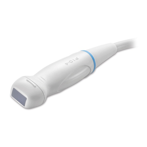 |
||||||||||||||||||||||
| Product Overview | The Veterinary Phased Array Cardiac Ultrasound Probe is designed for veterinary echocardiography and cardiovascular examinations requiring access through narrow acoustic windows. Its compact phased array footprint produces a sector-shaped ultrasound image, making it particularly suitable for cardiac imaging between ribs. The probe can be used with compatible veterinary ultrasound systems for assessment of cardiac structure, chamber dimensions, valve motion, myocardial movement and blood-flow-related examinations when supported by the host ultrasound platform. It is suitable for veterinary hospitals, companion animal clinics, equine practices, breeding centers, research facilities and professional livestock veterinary services. IMV Technologies lists phased array probes among the compatible transducers for its ExaPad veterinary ultrasound platform, which supports cardiac imaging as well as equine and farm animal applications. :contentReference[oaicite:0]{index=0} |
||||||||||||||||||||||
| Key Features | Phased Array Technology: A compact transducer footprint generates a sector-shaped image suitable for imaging through restricted intercostal spaces. Cardiac Imaging Application: Designed for veterinary echocardiography, including visualization of cardiac chambers, walls, valves and associated structures. Small Acoustic Window: The compact scanning surface helps improve probe positioning between ribs, which is important for thoracic and cardiac examinations. Real-Time Dynamic Imaging: Supports observation of rapid cardiac movement when paired with a compatible veterinary ultrasound scanner. Veterinary-Oriented Ergonomics: Handheld configuration is designed for stable positioning during repeated examinations. Flexible Equipment Matching: Probe frequency, connector type and compatible ultrasound system can be supplied according to project requirements. |
||||||||||||||||||||||
| Structural Components | Phased Array Transducer: Multiple piezoelectric elements are electronically controlled to steer and focus the ultrasound beam. Compact Acoustic Head: Designed for access through narrow intercostal spaces during veterinary cardiac scanning. Acoustic Lens: Provides the ultrasound contact surface used together with suitable coupling gel. Ergonomic Probe Housing: Protects the internal transducer assembly and supports controlled hand positioning. Flexible Signal Cable: Transfers imaging signals between the probe and ultrasound scanner. Ultrasound Connector: Connector configuration is matched to the intended veterinary ultrasound platform. |
||||||||||||||||||||||
| Working Principle | A phased array probe uses a group of small transducer elements that are activated with precisely controlled timing differences. These timing delays electronically steer and focus the ultrasound beam without requiring mechanical movement of the probe head. The resulting sector-shaped field of view starts from a relatively small contact area and expands deeper into the tissue. This scanning geometry is well suited to veterinary echocardiography because the probe can be positioned between ribs while providing a broader image of the heart at greater depth. Echoes returning from cardiac tissues are processed by the ultrasound system to generate real-time images. When the compatible scanner supports Doppler functions, the same probe may also be used for blood-flow assessment according to the system configuration. |
||||||||||||||||||||||
| Technical Specifications |
|
||||||||||||||||||||||
| Applications | Veterinary Echocardiography: Used for real-time examination of cardiac chambers, myocardium, valves and surrounding structures. Companion Animal Cardiology: Suitable for cardiac examinations in dogs and cats when matched with the appropriate frequency and ultrasound system. Equine Cardiac Examination: Can be used with compatible veterinary ultrasound platforms for selected equine cardiology applications. Livestock Veterinary Services: Applicable to selected cattle, goat-farming-equipment/" target="_blank" class="keylink">sheep, goat and other farm animal cardiac examinations when appropriate probe configuration and examination protocols are used. Cardiac Motion Assessment: Supports visualization of dynamic myocardial and valve movement. Doppler Examination: When connected to an ultrasound scanner equipped with Doppler functions, it may support evaluation of blood-flow direction and velocity. |
||||||||||||||||||||||
| Customization Options | Probe specifications can be matched to different veterinary ultrasound platforms and animal examination requirements. Available options may include: • Frequency configuration according to animal size and examination depth • Connector type according to ultrasound scanner model • Cable configuration available upon request • Probe configuration for companion animals, horses or livestock • OEM and project procurement options • Packaging configuration according to distributor requirements Customers should provide the ultrasound scanner brand, model, intended animal species and clinical application before ordering so that compatibility can be confirmed. |
||||||||||||||||||||||
| Installation & Maintenance | Confirm probe compatibility with the veterinary ultrasound scanner before installation. Connect the transducer according to the ultrasound system manufacturer's operating procedure. After startup, select the appropriate cardiac preset and probe configuration. Apply suitable ultrasound coupling gel to the examination area to provide effective acoustic contact. For thoracic examinations, position the probe carefully within the appropriate intercostal acoustic window. Avoid dropping the probe, striking the acoustic surface, pulling the cable or repeatedly bending the cable close to the probe housing or connector. After examination, remove residual coupling gel and clean the probe using a compatible veterinary ultrasound transducer cleaning method. Do not immerse the connector or other non-immersible components unless specifically approved by the equipment manufacturer. |
||||||||||||||||||||||
| Project Cases | The phased array cardiac probe can be supplied as part of veterinary ultrasound equipment packages for animal hospitals, specialist cardiology clinics, equine practices, livestock veterinary teams, breeding centers, distributors and agricultural equipment procurement projects. For mixed-practice and equine projects, the probe can be combined with compatible portable ultrasound platforms that support multiple transducer types. IMV Technologies identifies cardiac imaging as one of the clinical applications of its ExaPad system and lists compatible phased array probes within its veterinary ultrasound probe range. :contentReference[oaicite:1]{index=1} |
||||||||||||||||||||||
| Product FAQ | Q: Why is a phased array probe suitable for cardiac ultrasound? A: Its small acoustic footprint can fit between ribs while producing a wider sector-shaped image at depth, which makes it suitable for echocardiographic examinations. Q: Can this probe be used for dogs and cats? A: Yes. Phased array probes are commonly used for companion animal cardiology. The appropriate frequency and probe model should be selected according to animal size and required imaging depth. Q: Can it be used for horses? A: Yes, when the probe frequency and compatible ultrasound platform are suitable for equine cardiac imaging. IMV Technologies lists phased array probe compatibility within its equine ExaPad ultrasound range. :contentReference[oaicite:2]{index=2} Q: Does the probe support Color Doppler? A: Doppler capability depends on the connected ultrasound scanner. Compatible systems equipped with Color Doppler, Pulse Wave Doppler or other cardiac functions may use the probe for corresponding examinations. Q: Can it connect to any veterinary ultrasound system? A: No. Connector interfaces, supported probe electronics and frequency ranges differ between ultrasound systems. Compatibility should be confirmed before purchase. Q: Can the probe configuration be customized? A: Frequency, connector configuration, cable options and project packaging can be supplied according to project requirements and compatible equipment. |
||||||||||||||||||||||
| Product Gallery |    |
- Home
-
Products
- Pig Farming Equipment
- Cattle Farming Equipment
- Sheep & Goat Farming Equipment
- Poultry Farming Equipment
- Feeding & Drinking Systems
- Veterinary & Breeding Equipment
- Testing & Diagnostic Equipment
- Cleaning & Biosecurity Equipment
- Climate Control Equipment
- Livestock Weighing Equipment
- Manure & Waste Management
- Farm Transport & Handling
-
Solutions
- Pig Farm Solutions
- Dairy Farm Solutions
- Beef Cattle Farm Solutions
- Sheep & Goat Farm Solutions
- Layer Farm Solutions
- Broiler Farm Solutions
- Feeding & Drinking System Solutions
- Climate Control Solutions
- Biosecurity & Disinfection Solutions
- Manure & Waste Management
- Smart Farming Solutions
- Farm Renovation and Upgrade Solutions
- Partners
- Projects
- Resources
- News
- About Us
- Contact

Comments & Inquiries
Leave your questions or requirements below. We will reply as soon as possible.